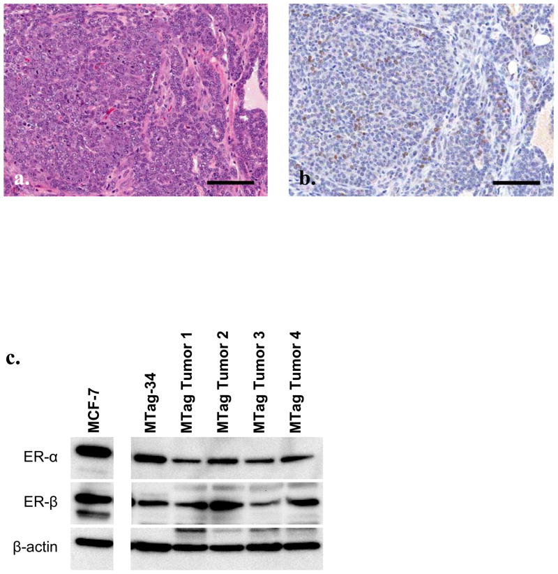
Figure 1.
MTag.Tg tumors and cell line express estrogen receptor. Representative Mtag.Tg tumor stained for H&E (a) and ER-α (b). Black bar indicates 100 μm. (c) Western blot for ER-α and β in MTag.Tg tumors and derived cell line lysates. MCF-7 cell lysates serves as a positive control, β actin as loading control.

