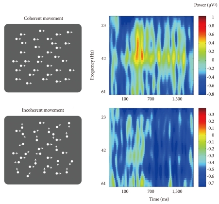
Figure 2.
Increase in gamma activity during the perception of coherent and incoherent dot motions by healthy subjects. The left column shows the two stimulus conditions, for coherently displaced dots from left to right (upper) and for incoherently displaced dots at randomly generated angles (lower). The right column shows time-frequency spectrograms of averaged electroencephalography power for coherent (upper) and incoherent (lower) dot motions in the channel Pz (Figure courtesy of Giri P. Krishnan).

