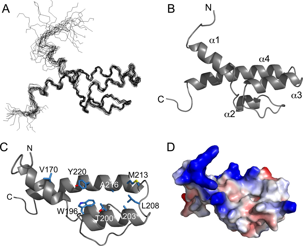
Fig 2. Solution Structure of Gal11-ABD1.
(A) NMR ensemble of 20 low-energy Gal11-ABD1 structures. Average pairwise RMSDs for the ordered backbone atoms of residues 163–187, 191–193, 195–232 is 0.9Å. (B) Ribbon representation of the Gal11-ABD1. (C) Orientation from (A) was rotated ~90 degrees about the x-axis to highlight the residues from α1, α3, and α4 that form the ABD1 hydrophobic cleft (shown in stick representation with carbons in blue, oxygens in red, and sulfur in yellow. (D) The surface electrostatic potential of ABD1 oriented as in (B). Red, negatively polarized; blue, positive; white, non-polar.

