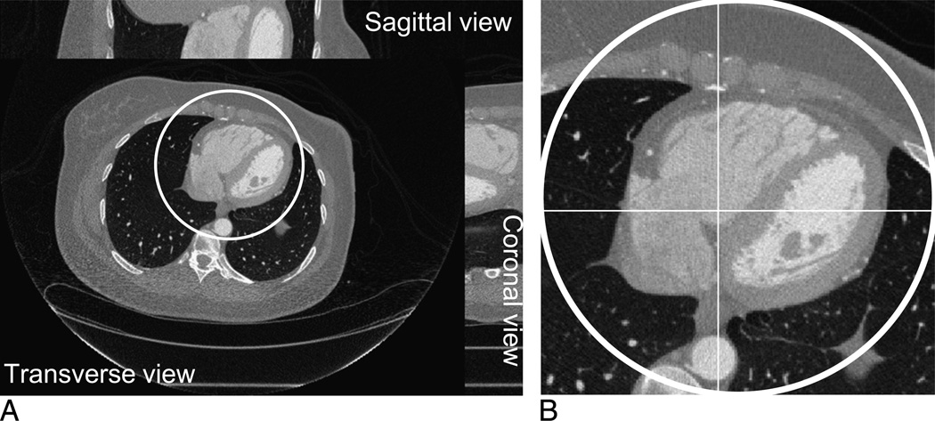
FIGURE 2.
A volumetric image reconstructed by the conventional filtered back projection algorithm from one helical turn (64 slices) clinical patient data set collected on a GE Discovery CT750 HD scanner. A, Transverse, sagittal, and coronal slices of the image volume. B, The magnified interior cardiac region of the transverse slice no. 32.

