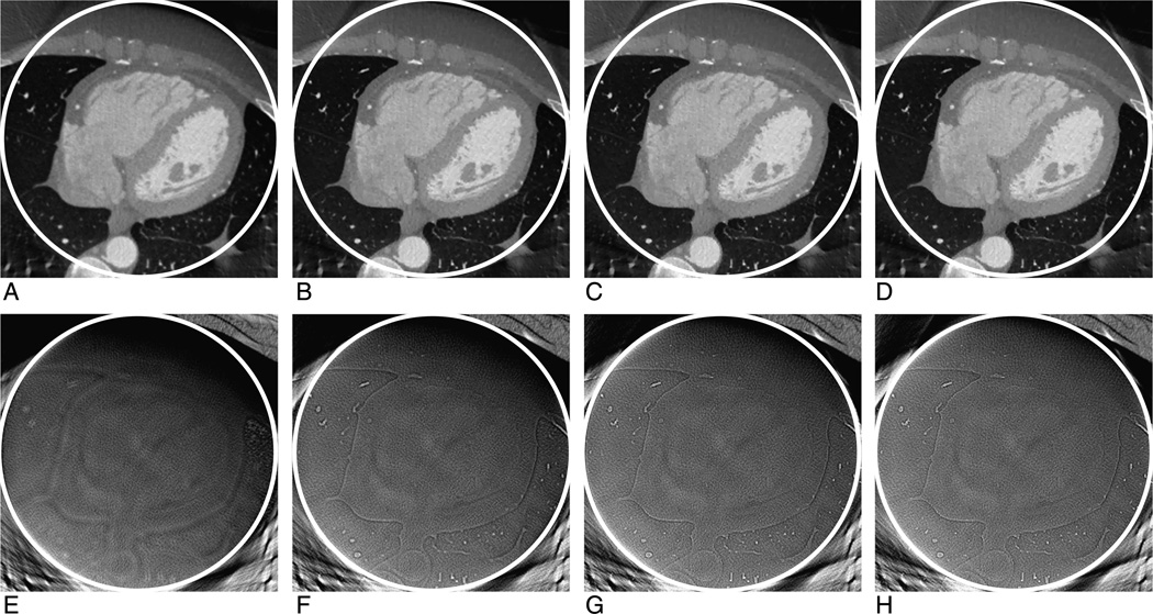
FIGURE 3.
Compressive sensing–based interior reconstruction of a cardiac region from a clinical patient data set collected on a GE Discovery CT750 HD scanner. From left to right, the iteration number for the top row images (Window Width, W = 2000 HU; Window Center, C = 0 HU) are 5, 10, 15, and 20, respectively. The bottom row images are the corresponding difference images between the interior reconstruction and global reconstruction (Window Width, W = 200 HU; Window Center, C = 0 HU).

