Figure 8.
Localization of UGT76B1 Expression Using UGT76B1pro:GUS-GFP Lines.
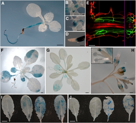
Transgenic plants harboring UGT76B1pro:GUS-GFP constructs were stained for GUS activity in different developmental stages ([A] to [D] and [F] to [J]) or examined for GFP fluorescence by confocal microscopy (E) (see Methods). Results were consistent among at least two independent transgenic lines.
(A) to (D) Eight-day-old seedling (A) with leaf primordia (B), leaf hydatodes (C), and root tip (D).
(E) Lateral root of a 1-week-old seedling grown on an agar plate. Cell walls were counterstained with propidium iodide. The red and blue lines indicate the positions where the vertical (right part) and longitudinal (left part) optical cross sections were taken, respectively. Vertical and longitudinal projections are separated by the purple line.
(F) A 17-d-old plant.
(G) A 28-d-old plant.
(H) Inflorescence of a 36-d-old plant. The inset shows a magnification of the stigma and anthers.
(I) Two leaves of 5-week-old plants 8 h after mock treatment (left) or after inoculation with Ps-avir (right).
(J) Two leaves before (left) or 6 h after (right) mechanical wounding using a forceps.
Bars = 1 mm in (A) and (F), 0.1 mm in (B) to (D), 30 μm in (E), 0.5 cm in (G), (I), and (J), and 0.5 mm in (H).

