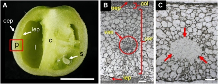
Laser capture microdissection of tomato fruit. Tomato fruit in cross section (A). Labels indicate pericarp (p), empty locule (l), columella (c), seed (s), outer epidermis (oep), and inner epidermis (iep). Cryosection of the tomato fruit pericarp before microdissection (B). Labels indicate outer epidermis (oep), collenchyma (col), vascular bundle (vas), parenchyma (par), and inner epidermis (iep) of the pericarp. Cryosection after microdissection of the vascular bundle; dissected area is indicated by arrows (C). Bars = 5 mm in (A) and 100 μm in (B) and (C). (Reprinted from Figures 1A, 1D, and 1G of Matas et al. [2011].)

An official website of the United States government
Here's how you know
Official websites use .gov
A
.gov website belongs to an official
government organization in the United States.
Secure .gov websites use HTTPS
A lock (
) or https:// means you've safely
connected to the .gov website. Share sensitive
information only on official, secure websites.
