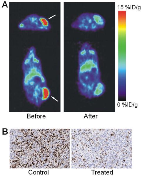
Fig. (4).
PET imaging of VEGF expression. (A) Transversal (top) and coronal (bottom) PET images of 89Zr-bevacizumab in xenograft mice before (left) and after (right) treatment with a Hsp90 inhibitor. Arrows indicate the tumor. (B) Ki67 staining of the tumor tissue corroborated the PET results. Adapted from [58].

