Figure 5.
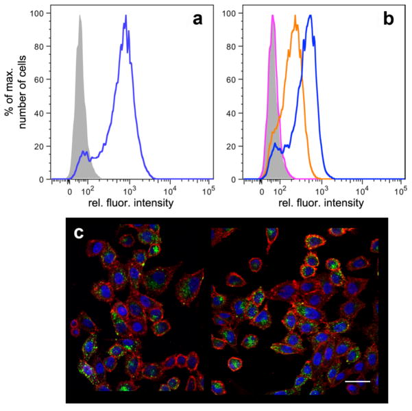
Packaged GFP-dependent analysis of cell binding and internalization. a) Flow cytometry of CD22-CHO cells treated with buffer alone (grey) or fluorescently labeled anti-CD22 antibody (blue); 4°C for 1 h, followed by washing. b) As in panel (a); cells treated with buffer (grey), 5 (25 μg/mL, 10 nM in particles, pink), 6 (2.5 μg/mL, 1 nM in particles, orange), or 6 (25 μg/mL, 10 nM in particles, blue). c) Representative confocal laser microscopy image of CD22-CHO cells treated with 6 for 1 h at 37°C. Blue = DAPI stained nuclei, red = cell membrane (wheat germ agglutinin AlexaFluor® 555 conjugate), green = encapsidated GFP, scale bar = 30 μm. Negative control images showing no detectable binding or internalization of GFP-containing particles in the absence of CD22 on the cells or ligand 2 on the particles are included in Supporting Information.

