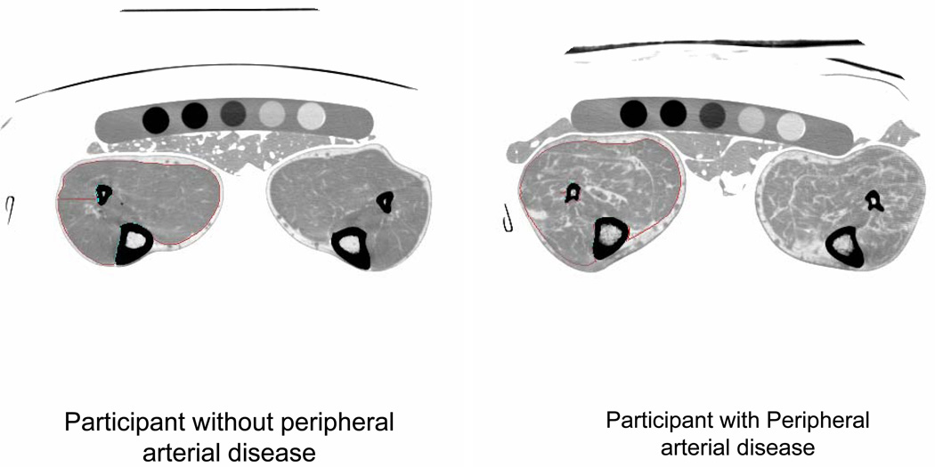
Figure. Calf images from a participant with and without peripheral arterial disease.*.
* A phantom and gel packs are shown below the legs. A marker indicates the right lower extremity. In this example, the right calf muscle in each participant is outlined with specially designed software, allowing quantification of calf muscle characteristics.

