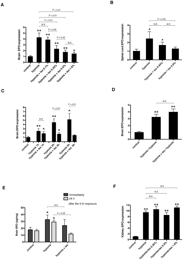
Figure 1. Effect of isoflurane on EPO expression in mouse CNS and kidney.
(A, B, and F) Six-week-old BALB/c mice were exposed to 10% O2 (hypoxia) in the presence of various concentrations of isoflurane for 3 hours (n = 6–15), or (C) exposed to 10% O2 (hypoxia) with 0.5% isoflurane for the indicated periods of time. (D) 24 hours after the hypoxic exposure with or without 0.5% isoflurane, 6-week-old BALB/c mice were re-exposed to hypoxia (10% O2) for 3 hours. EPO mRNA in the brain (A, C, and D), spinal cord (B) and kidney (F) was assayed with real-time RT-PCR analysis. (E) Immediately or 24 hours after the 5-hour hypoxic (10% O2) exposure with or without 0.5% isoflurane, EPO protein concentration (pg/ml) in the brain was quantified with ELISA and divided by the total protein concentration (mg/ml) of each mouse brain. Number of animals per treatment conditions is 6 (C–E). Data are presented as mean ± SD. The expression levels of EPO were normalized to that of 18S and expressed relative to the mean of control mice (A, B, C, D and F). *P<0.05, **P<0.01 versus control, N.S.; not significant (Mann-Whitney U-test).

