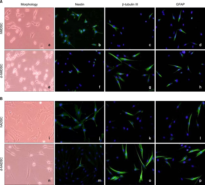
FIG. 1.
Morphology and immunocytochemistry of nestin, β-tubulin III, and GFAP before and after human muscle-derived stem cells (hMDSCs) and human-adipose tissue derived stem cells (hADSCs) differentiation. Morphology and double fluorescent immunocytochemistry for nestin (green), β-tubulin III (green), and GFAP (green) shown in (A) hMDSCs, differentiated hMDSCs (d-hMDSCs), and (B) hADSCs, differentiated hADSCs (d-hADSCs). Nuclei are stained with DAPI (blue). Magnification is ×100.

