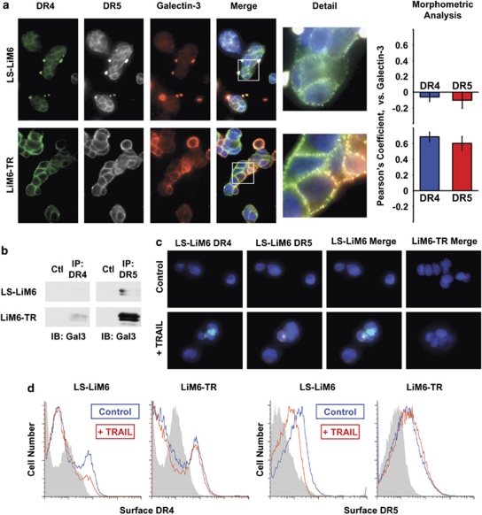
Figure 6.
Surface galectin-3 anchors DR4 and DR5 in nano-clusters and impedes endocytosis. (a) Cells were labeled with anti-DR4 (green), anti-DR5 (gray), anti-galectin-3 (red) and nuclear staining by DAPI (blue), and surface labeling was visualized with Alexa Fluor-conjugated secondary antibodies by confocal microscopy. The white boxes of merged images are magnified to show detailed morphology (detail). Morphometric analysis performed by the 3I's Slide book 5.0, computing Pearson's coefficient for degree of overlapping. (b) Lysates from LS-LIM6 or LIM6-TR cells were immunoprecipitated with either anti-DR4, anti-DR5, or control IgG antibodies. The precipitates were immunoblotted with anti-galectin-3 antibody (TIB 166). (c) TRAIL-dependent internalization of DR4 and DR5. Cells were pre-labeled with anti-DR4 (green) and anti-DR5 (gray) antibodies, and exposed to 100 ng/ml TRAIL on ice for 1 h. The temperature was raised to 37°C for 30 min to allow receptor internalization or kept at 0°C for controls. Surface labeling was removed by treatment with 2 m acetic acid, then the cells were fixed and permeabilized. Internalization of death receptor immunocomplexes was visualized with Alexa Fluor-conjugated secondary antibodies by confocal microscopy. (d) TRAIL-dependent loss of cell surface DR4 and DR5. Cells were treated with 100 ng/ml TRAIL for 30 min at 37°C followed by labeling with either anti-DR4 or anti-DR-5 antibodies and cell surface death receptor expression was determined by flow cytometry analysis

