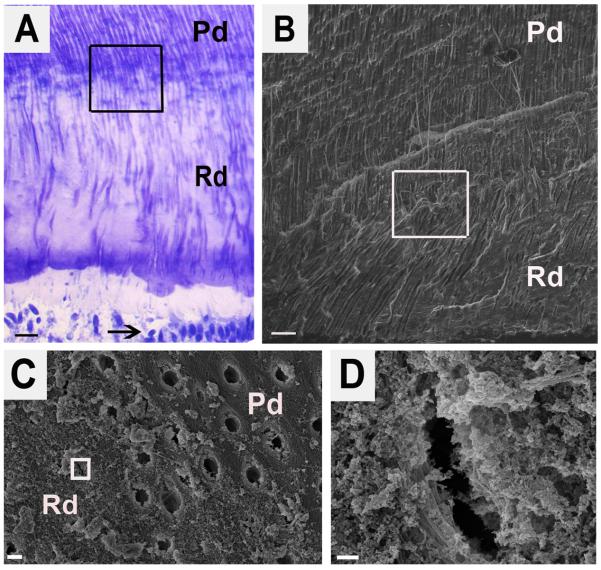
Figure 1.
Structure of reactionary dentin demonstrating fewer tubules that are more irregular and constricted. (A) Toluidine blue stain of longitudinal section of reactionary dentin. Pd and Rd indicate the area of physiologic dentin and reactionary dentin, respectively. Arrow points to odontoblasts. The boxed area encompasses the boundary between Pd and Rd. Scale bar 20 μm. (B) Scanning electron micrograph (SE detector at 10.00 kV, X500) of the boxed area from A. Scale bar 20 μm. (C) Scanning electron micrograph (SE detector at 10.00 kV, X7,500) of a cross-section of the boxed area from B and showing the boundary between Pd and Rd. Scale bar 2 μm. (D) A high magnification view (SE detector at 10.00 kV, X60,000) of the boxed area from C showing the morphology of a constricted tubule in the Rd. Scale bar 200 nm.

