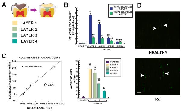
Figure 5.
Data demonstrating the gelatinase activity of MMP-2 in each dentin layer. (A) Diagram of dentin layers; layer 1 the superficial soft carious lesion, layer 2 inner soft carious lesion, layer 3 caries-affected dentin and layer 4 sound dentin beneath carious lesion including Rd. (B) Bar charts demonstrating gelatinolytic activity from extracted total dentin protein from each dentin layer. Gelatinolytic activity detected in Rd (layer 4) was almost entirely contributed by MMP-2. The activity was 5 times higher than gelatinolytic activity detected in the corresponding layer of dentin from healthy teeth. (C) MMP-2 activity in each dentin layer was calculated from a collagenase standard curve of the gelatin-fluorescein conjugate cleaved by Clostridium histolyticum. MMP-2 activity in the Rd (layer 4) was significantly higher than the healthy sample. All values depict means ± SD (n=14). * P≤ 0.05; ** P≤ 0.02. (D) Demonstration of MMP-2 expression in dentinal tubules in the Rd area by immunofluorescence on a semi-thin resin section. Positive labelling is indicated by bright green fluorescence. Staining for MMP-2 (arrowheads) observed in some dentinal tubules in the area of Rd was more abundant and intense compared to dentin from healthy teeth. Scale bar 20 μm.

