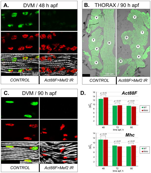
Fig. 5. Morphological and molecular effects of Mef2 silencing in post-fused IFM fibers.
A: Expression of MEF2 (green) in the nuclei (red), as well as nascent myofibrils (gray), in longitudinally sectioned DVM fibers shortly after completion of myoblast fusion in control and Act88F-Gal4-induced RNAi (Act88F>Mef2 IR5039) pupae at 48 h APF. B: General muscle localization in the thorax of control and RNAi pharate adults at 90 h APF, visualized by phalloidin staining (green). The DLM fibers (D) in RNAi animals (Act88F>Mef2 IR5039) are collapsed. The positions of other major thoracic muscles, DVMs (V) and TDT (T), remain unchanged. C: Longitudinally sectioned DVMs, stained for MEF2 (green), nuclei (red), and myofibrils (gray) in control and RNAi pharate adults (Act88F>Mef2 IR5039) at 90 h APF. Lack of MEF2 results in a wavy arrangement of myofibrils, although their structure is similar to control. D: Comparative analysis of expression of Act88F (upper panel) and Mhc (lower panel) between control (green bars, WT) and Act88F>Mef2 IR5039 (red bars, RNAi) samples, obtained from IFMs (combined DVM and DLM fibers) at 48 h, 72 h, and 90 h APF. Realtime qRT-PCR data are shown as the lag in detection (ΔCt) between transcripts of interest, and 18S and 28S rRNA, used as internal references (details are in Material and Methods) ± s.d. Student’s t-test p value > 0.05 indicates statistically non-significant differences. Note that the expression of Act88F lags slightly in the RNAi samples at the 48 h APF time-point, but not at any other stages. Mhc transcript levels are not significantly different between control and knockdown samples.

