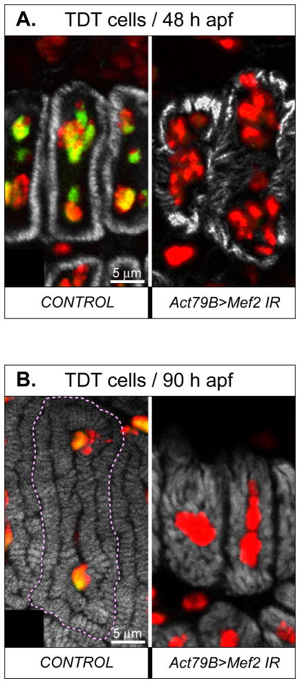
Fig. 8. Mef2 knockdown in the TDT does not prevent myofibrillogenesis, but affects organization of myofibril arrays and general myofiber morphology.
A, B: Transversely cut individual TDT muscle fibers at 48 h (A) and 90 h (B) APF, in control and RNAi (1151>Mef2 IR5039) pupae. The superimposed images of three fluorescent channels, corresponding to MEF2 (green), DNA (red), and F-acting (grey) are shown. The yellow color indicates an overlap between the green and the red signals. Note that nuclei of RNAi fibers contain only the red signal and are significantly disorganized. Fibrillar organization is disturbed in RNAi fibers at both time points, but the sizes of individual myofibrils have increased between 48 h and 90 h APF.

