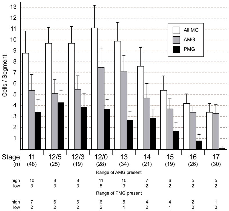
Fig. 2. Variation in the number of MG during embryogenesis.
Comparison of the number of AMG, PMG, and total MG from stages 11 to 17 from the analysis of stained confocal images. The number of segments examined (n) is noted below each stage. Data are means ± standard error of the means. At stages 11 to 12/0, all MG were counted using the expression of 12xSu(H)bs-lacZ and wrapper. AMG were high wrapper and low 12xSu(H)bs-lacZ and PMG were low wrapper and high 12xSu(H)bs-lacZ. From stages 13 to 17, MG were identified by position and expression of wrapper, (high in AMG and low in PMG). The numbers of AMG and PMG are relatively constant from stages 11 to 12/3. From stages 12/0 to 17, average AMG number spikes before declining to approximately 3 AMG/segment while the number of PMG declines to near 0 by stage 17. At the bottom, the range of values for the number of AMG or PMG at each stage is shown as the highest (high) and lowest (low) number counted.

