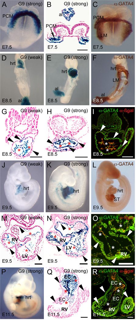
Fig. 2. TheGata4G9 enhancer is active in cardiac progenitors in the cardiac crescent and the linear heart tube and restricts to endocardium during mouse cardiogenesis.
X-gal stained embryos photographed as whole-mounts (A, D, E, J, K, P) or following transverse sectioning of stained embryos (B, G, H, M, N, Q) are shown. For comparison, wild type embryos stained as whole mounts with anti-GATA4 antibody are shown (C, F, L). In addition, transverse frozen sections stained for immunofluorescence with anti-p-galactosidase and anti-GATA4 antibodies are also shown (I, O, R). Strong Gata4-G9-lacZ transgenic lines are pictured in B, E, H, I, K, N, and O; weak Gata4-G9-lacZ lines are pictured in D, G, J, M. Asterisks mark endocardium and arrowheads mark myocardium. al, allantois; EC, endocardial cushions; hrt, heart; LM, lateral mesoderm; LV, left ventricle; PCM, precardiac mesoderm; RV, right ventricle; ST, septum transversum. Bars in all panels = 100 µM.

