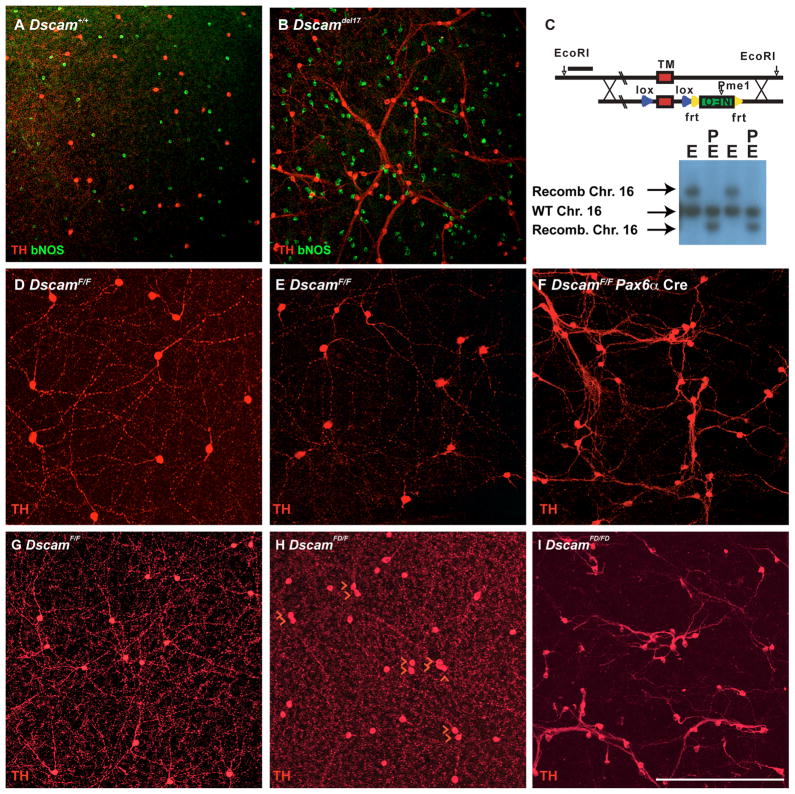
Figure 1. DSCAM-deficient phenotypes and Dscam conditional allele.
A and B, Retinas stained with antibodies to tyrosine hydroxylase (TH) and brain nitric oxide synthetase (bNOS) to label dopaminergic (DA) and bNOS-positive amacrine cells, respectively. DA cell or bNOS cell neurites fasciculate with the neurites of other cells of the same type in Dscamdel17/del17 retinas but not across cell types. C, A conditional allele of Dscam was generated by flanking the exon encoding the Dscam transmembrane domain with loxP sites. An upstream EcoRI site was used for identifying homologous recombinants by Southern blotting. Recombination resulted in a 2 kb upshift when digested with EcoRI (E), or a 2 kb decrease in size if double digested with EcoRI (E) and PmeI (P), which cuts in the Neo-selectable marker. The results of Southern blotting DNA preps for two independent ES cell colonies are shown. The wild type Chromosome 16 (arrow) is cut by EcoRI resulting in a single band of approximately 12 kb. The recombined Chromosome 16 (arrow) is cut by EcoRI resulting in a single band of approximately 14 kb. The wild type Chromosome 16 (arrow) is cut by EcoRI and PmeI resulting in a single band of approximately 12 kb (PmeI has no cut sites within the EcoRI fragment). The recombined Chromosome 16 (arrow) is cut by EcoRI and PmeI resulting in a single band of approximately 10 kb (recombined sequence adds a PmeI site within the EcoRI fragment). D–F, Dscam+/+, DscamF/F and DscamF/F Pax6α Cre retinas were collected at postnatal day 15 (P15) and stained with an antibody to TH. Cell bodies of wild type and DscamF/F DA cells are spaced and have well arborized neurites, whereas conditional deletion of the Dscam transmembrane domain with Pax6α Cre results in clumped DA cells and neurites. G–I, Germ line deletion of the conditional allele (DscamFD). Dopaminergic amacrine (DA) cells in retinas from P28 mice were stained with antibodies to TH. The number of DA cells directly adjacent to other DA cells was counted in each of four retinas and compared using a T-Test. DscamF/F retinas had 10.5 +/− 1 DA cell pairs/retina while DscamFD/FD retinas had 30.75 +/− 5.1 pairs/retina: T-Test=0.001. The scale bar in I is equivalent to 538.2 μm in A and B, 387.5 μm in D–F and 228 μm in G–I.

