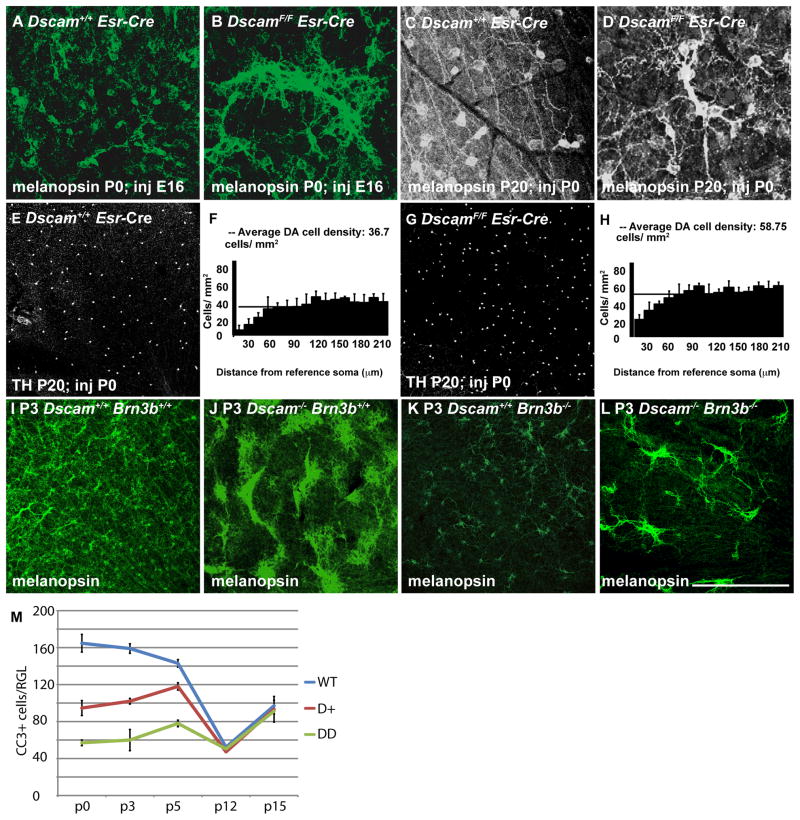
Figure 2. Independence of Dscam-dependent arborization and cell number regulation.
A–H, The Dscam conditional allele was targeted using a tamoxifen-inducible Esr Cre. A and B, Retinas from P0 Dscam+/+ Esr Cre and DscamF/F Esr Cre mice, in which induction of Esr Cre was performed at embryonic day 16 (E16), were stained with antibodies to melanopsin. Widespread fasciculation and aggregation of mRGCs was observed in DscamF/F Esr Cre retinas compared to Dscam+/+ Esr Cre retinas (P0; N=4). C–D, Retinas from P20 Dscam+/+ Esr Cre and DscamF/F Esr Cre mice, in which induction of Esr Cre was performed at P0, stained with antibodies to melanopsin (N=3 and 5, respectively). Dscam+/+ Esr Cre and the majority of DscamF/F Esr Cre mRGCs had arborized dendrites and spaced soma. Occasional examples of fasciculation were observed in the DscamF/F retinas. E and G, Retinas from P20 Dscam+/+ Esr Cre and DscamF/F Esr Cre mice, in which induction of Esr Cre was performed at P0, stained with antibodies to tyrosine hydroxylase (TH) (N=3). F and H, Density Recovery Profile (DRP) analysis was performed on DA cells from mice in which Esr Cre was induced at P0. The horizontal bar running across the vertical columns represents the average cell density. Vertical columns indicate the average number of cells located at a given distance from other cells of the same type, termed the reference soma. F, DA cells in retinas from Dscam+/+ mice had an average cell density of 36.7 and had an exclusion zone, indicated by the lack of DA cells in close proximity to other DA cells, as represented by the vertical columns on the left hand side of the chart with reduced values compared to the average cell density. H, DA cells in retinas from DscamF/F mice had an average cell density of 60.4 and had an exclusion zone. I–L, Retinas from postnatal day 3 (P3) wild type, Dscamdel17/del17, Brn3b−/− and Dscamdel17/del17 Brn3b−/− mice were stained with antibodies to melanopsin, to label mRGCs. I, In the wild type retina mRGCs are distributed across the retina (N=4). J, In the Dscamdel17/del17 retina mRGC somata are densely clumped (N=4). K, The number of mRGCs is greatly reduced in the Brn3b−/− retina and have spaced soma (N=4). L, The number of mRGCs is reduced in the Brn3b−/−;Dscamdel17/del17 retina. The remaining mRGCs present in the Dscamdel17/del17;Brn3b−/− retina are densely clumped (N=4). M, Whole retinas were stained with antibodies to cleaved caspase three (CC3) and the number of immuno-positive cells per retina was counted. A highly significant decrease in the number of cleaved caspase 3-postive neurons was observed when comparing Dscamdel17/del17, Dscamdel17/+ and Dscam+/+ at postnatal days 0, 3 and 5 (T-Test < 0.002). No differences were detected at postnatal days 12 and 15 except between heterozygous and wild type at p12, which was comparatively small (T-Test=0.015). The scale bar in (K) is equivalent to 194 μm in A and B, 127.8 μm in C and D, 775 μm in E and G and 387.5 μm in I–L.

