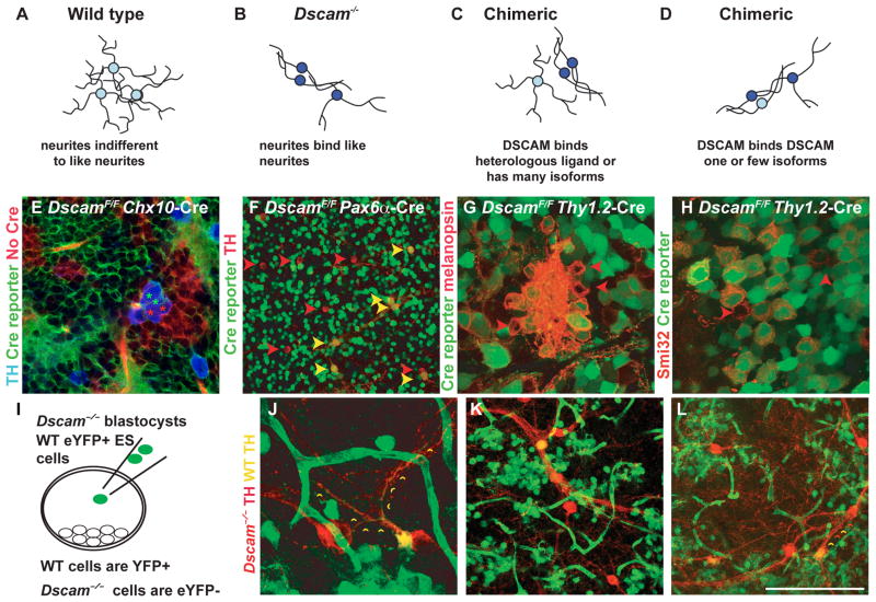
Figure 3. DSCAM-deficient cells induce mutant phenotype on wild type neighbors.
Potential mechanisms of DSCAM-activity. A, In the wild type retina many neurons of a given type (homotypic neurons) arborize dendrites in an overlapping fashion. B, In the Dscam-deficient retina, homotypic-neurites fasciculate with each other. C, If DSCAM activity in the retina is mediated through interactions with a heterologous ligand, wild type cells will be expected to maintain a wild type phenotype in a chimeric retina. D, If DSCAM activity in the retina is mediated through homophilic interactions, wild type cells are predicted to clump and fasciculate with juxtaposed Dscam mutant cells, but not other wild type cells. E, Chx10 Cre was used to delete Dscam from clonal columns of retina, Cre activity was monitored with a dual fluorescent Cre reporter such that dsRed (red) clonal columns lack Cre activity and eYFP (green) clonal columns indicate Cre activity (N>5). DscamFD/FD (green asterisk) and wild type (red asterisk), or multiple DscamFD/FD DA cells, were observed in aggregates as shown; however, multiple wild type DA cells were not observed to aggregate and fasciculate with each other. F, Image of central DscamF/F Pax6α Cre retina stained with antibodies to TH. Variable expression of Cre results in a mix of wild type and mutant DA cells. Mutant DA cells were found localized next to either other mutant DA cells or wild type cells but wild type cells were rarely found juxtaposed next to other wild type DA cells, as in the wild type retina. G and H, Dscam was deleted from a subset of RGCs using Cre under control of the Thy1.2 promoter, indicated by a Thy1-floxed-stop-YFP Cre reporter (N>3). Aggregates of melanopsin positive (G) and SMI-32 positive alpha RGCs (H) contain a mixture of wild type (YFP-negative) and deleted neurons (YFP-positive). I, GFP-positive ES cells that are wild type for Dscam were injected into Dscamdel17/del17 blastocysts to generate chimeric retinas. Dopaminergic amacrine cells within chimeric retinas were labeled with an antibody to TH (N=2). J–L, Wild type dopaminergic amacrine cells (GFP-positive; yellow arrow head) aggregated and fasciculated with mutant dopaminergic amacrine cells (GFP-negative; red arrowhead). Small yellow arrowheads are placed along an example of cofasciculating wild type and mutant DA cell neurites. The scale bar in (L) is equivalent to 194 μm in E and F, 95 μm in G, 70 μm in H, 90 μm in J and 132 μm in K and L.

