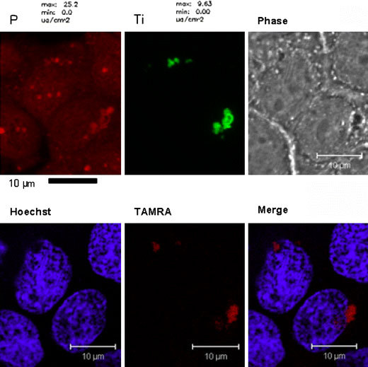
Figure 2.

Combining X-ray fluorescence microscopy and fluorescent confocal microscopy for the imaging of intracellular nanoconjugates. MCF-7 cells were transfected with TiO2-DNA nanoconjugates complimentary to genomic DNA encoding r18S rRNA. The DNA was fluorescently labeled with TAMRA. After treatment, cells were washed, fixed, and stained with Hoechst dye. Then they were analyzed by fluorescent confocal microscopy for the localization of TAMRA. Next, the same cells were dehydrated in 100% ethanol and analyzed at the 2-ID-D Beamline at the Advanced Photon Source at Argonne National Laboratories for the presence of titanium. Black bar scale represents 10 μm for XFM (top left and middle), and the white bar 10 μm for fluorescent confocal microscopy (top right, bottom row)
