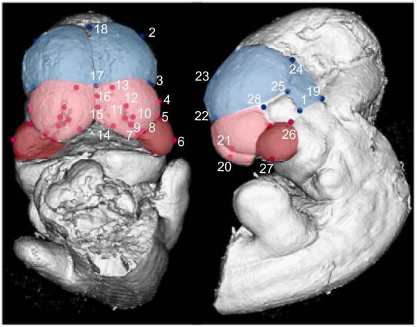
Figure 1.
Surface reconstruction of a micro-CT scan of an E10.5 C57BL6/J with landmarks listed on Tables 4-5. The blue region and landmarks are considered to be part of the forebrain; the red landmarks are part of the face; the pink area is the frontonasal process and the red area is the maxillary process.

