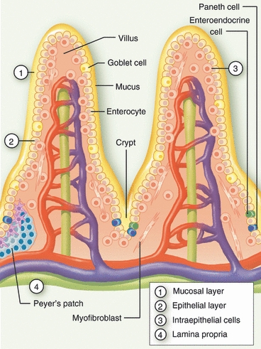
Figure 1.

Structural organization of intestinal mucosa. Schematic representation of the small intestinal mucosal barrier, consisting of a mucosal layer (1) and gradients of IgA and antimicrobial factors, the epithelial cells (2) made up of enterocytes, Paneth cells, goblet cells and enteroendocrine cells, intraepithelial lymphocytes (3), and the lamina propria (4). Additional secondary lymphoid structures, such as cryptopatches and Peyers’ patches are present in the lamina propria.
