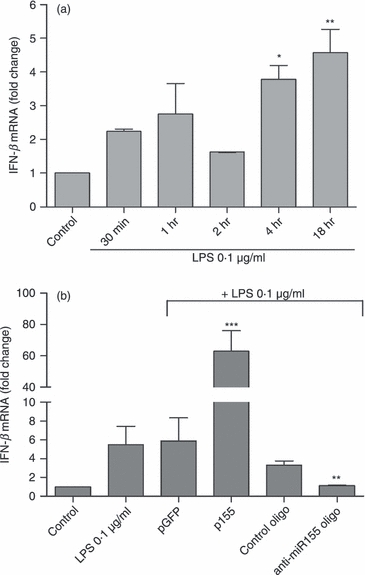
Figure 4.

Modulation of interferon-β (IFN-β) expression by microRNA-155 (miR-155). (a) N9 microglia cells were incubated with lipopolysaccharide (LPS) at 0.1 μg/ml for different periods of time (30 min, 1 hr, 2 hr, 4 hr and 18 hr). Results are presented as IFN-β mRNA fold change with respect to control (untreated cells). *P < 0.05 and **P < 0.01 compared with control. (b) The IFN-β expression levels were determined by quantitative real-time RT-PCR. N9 cells were transfected with anti-miR-155 oligonucleotides (anti-miR155 oligo) or miR-155-encoding plasmid (p155) complexed with cationic liposomes for 4 hr. Alternatively, N9 cells were transfected with non-targeting oligonucleotide (control oligo) or a plasmid encoding GFP (pGFP). Twenty-four hours after transfection, cells were incubated with LPS at 0.1 μg/ml for 18 hr and RNA extraction was performed. Results are expressed as IFN-β mRNA fold change with respect to control (untranfected and untreated cells). **P < 0.01 and ***P < 0.001 compared with LPS-treated cells in the absence of transfection. Results are representative of three independent experiments performed in triplicate.
