
Figure 3.
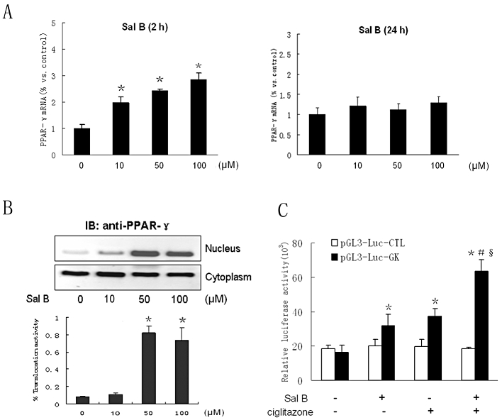
Effects of Sal B on PPARγ expression and transcriptional activity in h-monDC without ox-LDL stimulation. (A) Cultured h-monDC were treated with various concentration of Sal B for 2 h or 24 h. (B) The mRNA andprotein levels of PPARγ were examined by real-time RT-PCR and Western blot respectively. (C) pGL3-Luc-enhancer report vector with a GK promoter was transfected into h-monDC, and then the cells were treated with vehicle (PBS), Sal B (50 µM) and/or ciglitazone (25 µg·mL−1). Cell lysates were harvested, and both luciferase and β-galactosidase activities were analysed using the Dual-Luciferase reporter assay system. Data are shown as related luciferase activity in cells transfected with pGL3-Luc-GK vector or pGL3-Luc control vector after normalized to the β-galactosidase activity. Data were presented as means ± SD of four independent experiments. *P < 0.05, significantly different from control; #P < 0.05, significantly different from Sal B; §P < 0.05, significantly different from ciglitazone.