Figure 1.

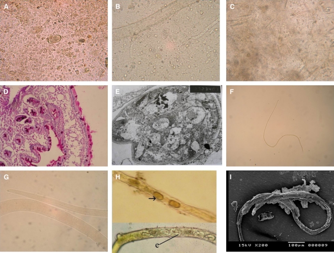
A, Translucent or yellowish, barrel-shaped, and thin-shelled Capillaria philippinensis eggs with rounded mucoid plugs protruding from both poles (×400). B, Adults, larvae, and distorted eggs in the stool of the same patient (×400). C, Charcot-Leyden crystals in the stool; they varied in number and size (×400). D, Jejunal biopsy showing numerous worm sections invading the mucosa (×400). E, Transmission electron microscopy of the jejunal mucosa showing the adult worm, which appears to lie in direct contact with an epithelial cell cytoplasm, with swollen mitochondria, distended rough endoplasmic reticulum, and atrophy of the villous surface of the intestinal wall. F, Capillaria philippinensis adult female; a: anterior end and p: posterior end (×40). G, Capillaria philippinensis esophagus; m: muscular portion and c: cellular portion (×400). H, The uterus contained either thin (e) or thick-shelled (arrow) eggs that were arranged in one row in the direction of the vulva (×200). I, SEM of an adult female with an envelope-like membrane covering most of the body surface.
