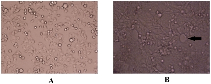
Figure 1. The cytopathic effects in HBECs after infection with RSV were observed under phase contrast microscopy.
A. normal HBECs; B. The classical pathologic changes of RSV-infected cells: enlarged fusion cell (×100). In this vision, variant shape, shrinkage, and enhanced refraction were present in cells, as well as enlarged fusion cells and intracytoplasmic eosinophilic inclusion, which were considered to be proof of RSV infection. A fusion cell is indicated by arrows.

