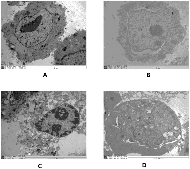
Figure 3. Subcellular structure of infected cells and subcellular localization of virus under electron microscopy.
A is normal HBECs (×10000); B is RSV-prolonged infected HBECs in early infection stage, fissure around nucleus is seen (×10000); C is also in early infection stage, expansion of endoplasmic reticulum and a large number of lysosomes in cytoplasm were present (×15000). Both cells in B and C had double nucleoli, which indicated the cell was proliferating rapidly; D is in late infection stage, enlarged fusion cells were present (×10000). Intracellular virus particles indicated by arrows were observed both in nucleus and endoplasmic reticulum.

