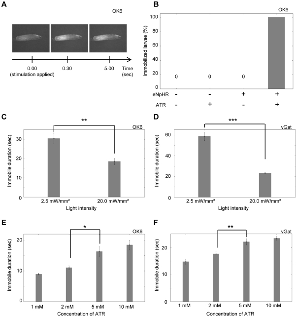
Figure 2. Optical inhibition of motor neurons.
Optical inhibition of motor neurons immobilized the larvae. OK6-Gal4 (A, B, C, E) and vGat-Gal4 (D, F) were used to express eNpHR in motor neurons. (A) Postures of a larva expressing eNpHR before (left) and after light stimulation (middle, 0.5 sec; right, 5 sec). The entire body was relaxed after light stimulation. (B) Dependence on the eNpHR transgene and ATR. Percentage of larvae immobilized over 5 sec in response to the continuous optical stimulation. n = 20 for each experiment. (C–F) Dependence on light intensity (C, D) and concentration of ATR (E, F). Average duration of the effective inhibition is plotted. n = 9∼10 for each experiment. Only a single stimulation was applied in this experiment and in Figs. 3 and 4. Thus, n represents the number of stimulation as well as the number of larvae examined. Error bars represent standard error. ***p<0.001, **p<0.01, *p<0.05; Student's t-test (C, D), ANOVA with Tukey-Kramer post-hoc test (E, F).

