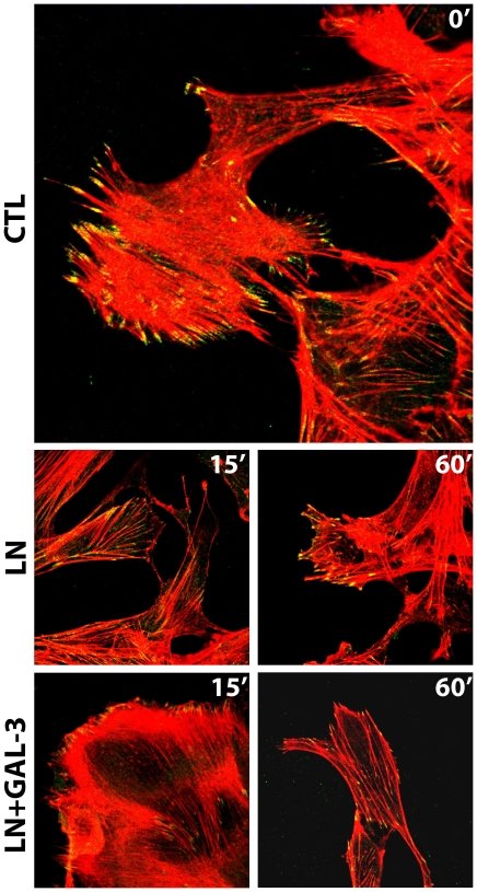
Figure 4. Extracellular galectin-3 (Gal-3) promotes the disassembly of stable focal adhesion plaques by decreasing the amount of phosphorylated FAK in the lamellipodia of migrating cells.
Σ12 cells were grown on coverslips and were subjected to the scrape assay either in the absence (ctl) or presence of laminin-111 (LN) or laminin-111 and 20 µg/mL galectin-3 (LN+gal-3) for 15 minutes. Cells were either fixed after 15 minutes after the migration stimulus. Confocal photomicrographs of typical fields are shown. Intracellular distribution of phosphorylated FAK (green) and organization of stress fibers (phalloidin staining, in red) are shown. Control cells showed mature adhesion plaques, as shown by staining of phosphorylated FAK. In the presence of galectin-3, there was a fast disassembly of the adhesion plaques indicated by the decrease of phosphorylated FAK in the lamellipodia.

