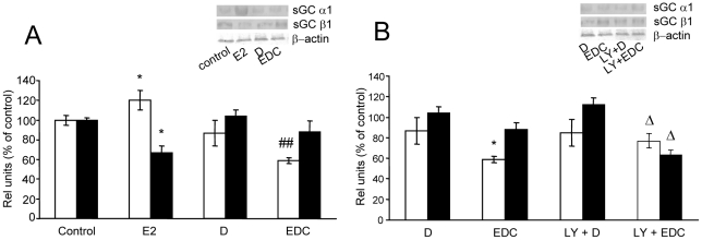
Figure 4. E2 decreased sGC α1 protein expression in a PI3k-dependent pathway involving non-nuclear ER but does not modify sGC β1 after 3 h of incubation.
Pituitary cells in culture were incubated with vehicle (control) or 1 nM estrogen dendrimer conjugate (EDC), unable to trespass cellular membrane for 3 h with or without 50 µM LY294002 (LY), a PI3K inhibitor, 30 min before treatment. Protein expression was evaluated by western blot. (A, B) Top, representative western blots. Bottom, Corresponding average densitometric values. Bars represent mean ± SE of α1 (open bars) and β1 (black bars) protein densitometric values normalized to β-actin, as percent of control (n = 3). ANOVA followed by Tukey's test, *P<0.05 vs. respective controls; ΔΔP<0.01 vs. E2; #P<0.05 vs. EDC.

