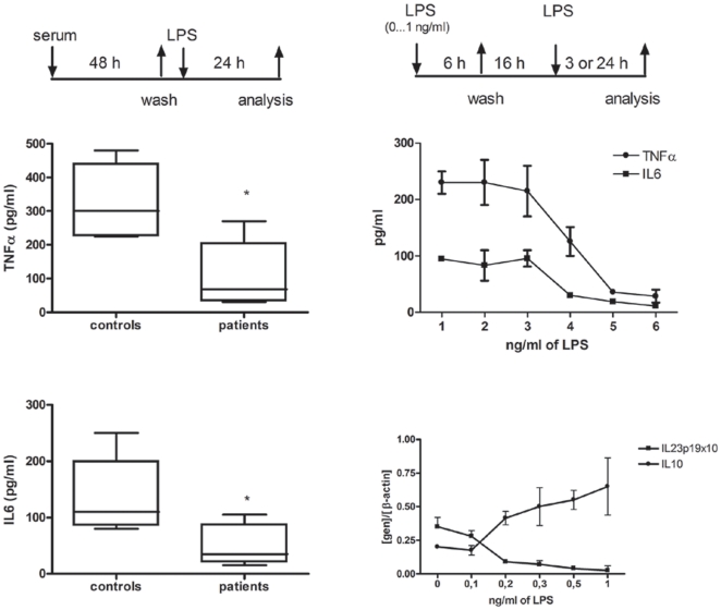
Figure 4. Concentrations of LPS detected in CF patients induce ET in healthy control M s.
s.
(A) Schematic representation of the employed experimental design. Monocytes, isolated from healthy controls, were cultured in presence of sera from CF patients (n = 14) and controls (n = 5) for 48 hours. Then, cultures were washed and fresh complete medium and LPS (10 ng/ml) added for 24 h. Next, TNFα (B) and IL6 (C) production were analyzed by ELISA in the supernatants. (D) Schematic representation of the employed endotoxin tolerance model (see Materials and Methods and ref. [10]). Cultures of monocytes, isolated from healthy controls, were pretreated with indicated LPS doses for 6 h. Then, cultures were washed and kept in complete medium for 16 h. After this period of recovery, the cultures were re-challenged with 10 ng/mL of LPS for 24 h (E) or 3 h (F). Next, TNFα and IL6 (E) production were analyzed by ELISA in the supernatants of the cultures and cells were harvested and mRNA of IL10 and IL23p19 were quantified by real time Q-PCR (F).

