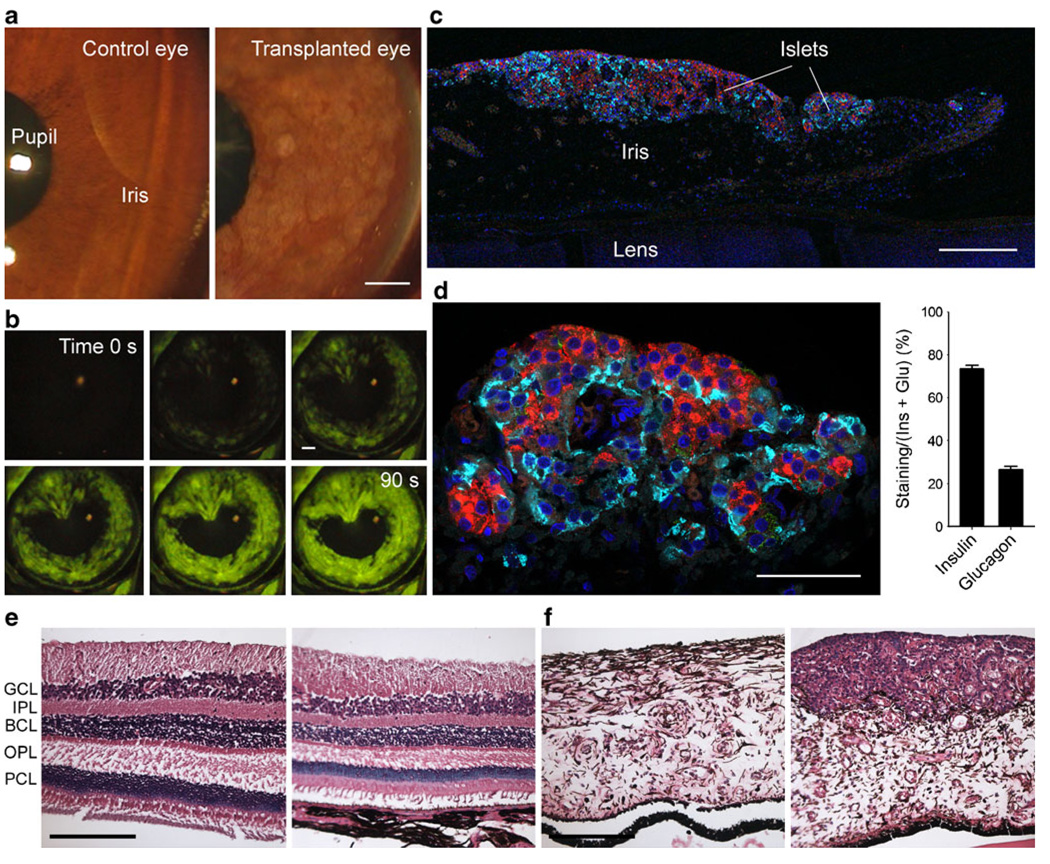
Fig. 1.
Pancreatic islets engrafted on the iris in the anterior chamber of the eye of a baboon model for type 1 diabetes. a Digital images of the anterior chamber of the eye of a baboon showing islet grafts on the iris (right). The anterior chamber and the cornea were as clear as those of the control, non-transplanted eye (left). In the transplanted eye, the angle was not obstructed by islets. b Fluorescein angiography performed at POD 24 revealed islet vascularisation. c Confocal image of a section of the anterior segment of the eye explanted at necropsy shows engraftment of islets on the iris. Insulin-labelled beta cells (red) and glucagon-labelled alpha cells (cyan) formed a distinct layer fully fused with the iris tissue. d The cytoarchitecture of intraocular islet grafts was preserved as seen by a typical ratio of beta cells to alpha cells (quantified on right panel as area of cell specific staining/[area of insulin staining+area of glucagon staining]×100, [n=47 sections]). e Vertical sections of the control, non-transplanted (left) and the transplanted eye (right) showing that the retina in the experimental eye is pristine (haematoxylin and eosin staining). BCL, bipolar cell layer; GCL, ganglion cell layer; IPL, inner plexiform layer; OPL, outer plexiform layer; PCL, photoreceptor cell layer. f Sections of the iris in the transplanted eye (right) showing islets engrafted on the iris, whose morphology is very similar to that of the iris of the control, nontransplanted eye (left) (haematoxylin and eosin staining). Scale bars, 200 µm (a–c, e, f) and 50 µm (d)

