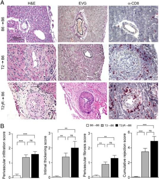
Figure 2.
Transplant vasculopathy induced by DC immunization. (A) Representative sections of transplanted B6→B6, T2→B6, and T2γR→B6 hearts on day 15 after DC immunization (H&E, EVG, and immunostaining for CD8). Original magnification ×400; scale bar, 100 μm. (B) Detailed analysis of immunopathological vascular alterations on day 15 post immunization. Individual scores for perivascular infiltration, intimal thickening, and perivascular fibrosis were determined, and the cumulative rejection score was calculated as the sum of the three individual parameters. Pooled data from four independent experiments (mean+SEM; B6→B6, n = 10; T2→B6, n = 18; T2γR→B6, n = 5). Statistical analysis was performed using one-way ANOVA with Bonferroni post test (***, p<0.001; **, p<0.01; ns, not significant).

