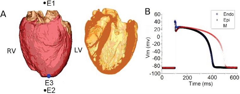
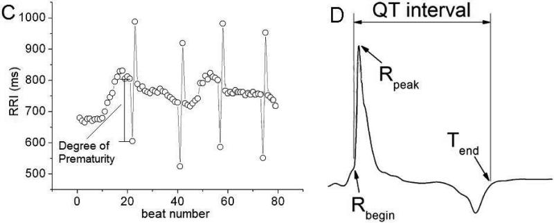
Figure 2.
A. Epicardial (left) and transmural (right) views of the biophysically-detailed MRI-based human ventricular model (at left, ventricles are in pink and atria in brown). Atria were insulated from the ventricles during the simulation. ECG electrodes are E1 and E2; pacing electrode is E3. B. Action potentials of human endo-, M, and epicardial cells. C. RRI sequence with PAs used as a pacing train in the electrophysiological simulations (see text for detail). D. ECG annotation and QT interval in one beat from a pseudo-ECG.


