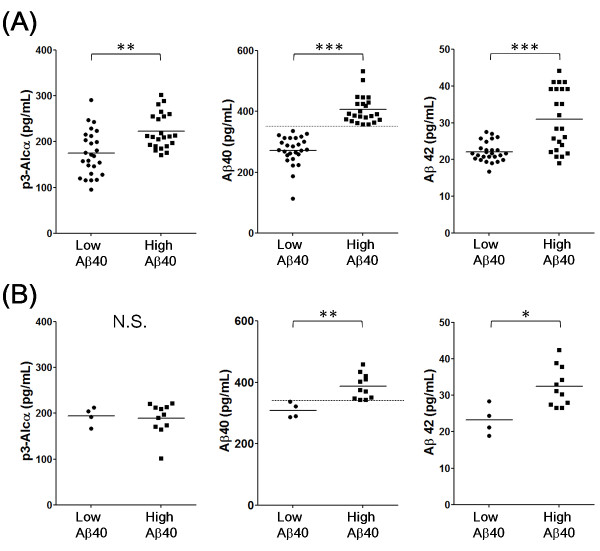
Figure 3.
Levels of p3-Alcα and Aβ in plasma from "low Aβ40" and "high Aβ40" populations of AD and FTLD subjects (Japanese cohort 1). Subjects with AD and FTLD were separated into two populations, who showed low (< 340 pg/mL) and high (> 340 pg/ml) Aβ40 levels (cut-off line is shown in the middle panels with dotted line). AD (A) and FTLD (B) are respectively indicated in two populations for p3-Alcα (left), Aβ40 (middle) and Aβ42 (right). Statistical analysis was performed using the Mann-Whitney U-test. *, P < 0.05; **, P < 0.01; ***, P < 0.001. N.S, not significant.

