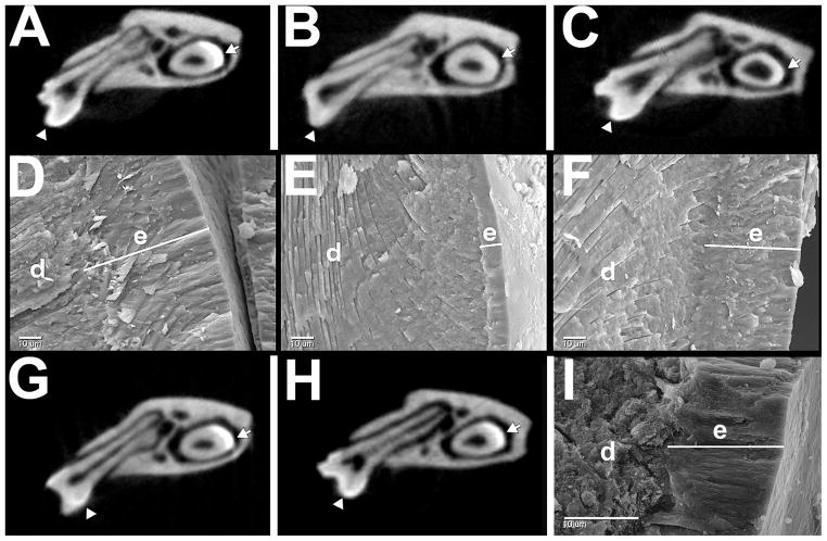
Fig. 3.
MicroCT and scanning electron microscopy analysis of enamel layers. A–C,G,H microCT at the level of the lower 1st molar; D–F,I scanning electron microscopy. Shown are wild-type (A,D); Amelx null (B,E); TgM180KO (C,F). TgM180 (G); TgP70T (H); (I) scanning electron microscopy of fractured surface of TgM180KO incisor enamel, which lacks substantial rescue as the transgene is expressed primarily in molar ameloblasts.

