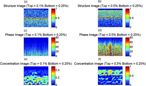
Figure 6.
Structure image for a phantom with a top layer ink concentration of (a) 0.1% and (b) 0.5%. Phase image for a phantom with a top layer ink concentration of (c) 0.1% and (d) 0.5%. Reconstructed concentration images for a phantom with a top layer ink concentration of (e) 0.1% and (f) 0.5%. The bottom layer ink concentration for both phantoms was 0.25%. White line of (f): 100 μm. The color bar of the structure image has arbitrary units, the color bar of the phase image has arbitrary units, and the color bar of the concentration image is in ink concentration.

