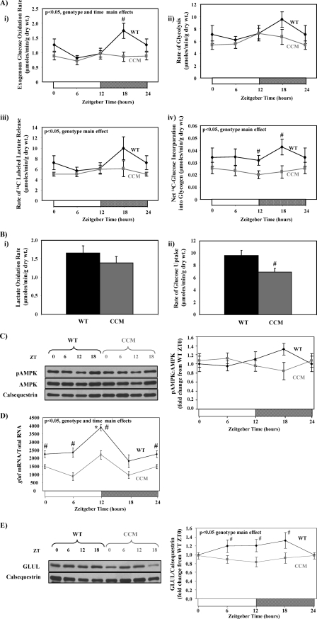
FIGURE 3.
Time-of-day- and cardiomyocyte circadian clock-dependent differences in parameters influencing entry of carbon into the HBP. Diurnal variations in rates of glucose oxidation (i), glycolysis (ii and iii), and glycogen synthesis (iv) in ex vivo perfused in WT versus CCM hearts are shown (A). Rates of lactate oxidation (i) and glucose uptake (ii) for WT versus CCM hearts at ZT18 are shown (B). Diurnal variations in P-AMPK levels in WT versus CCM hearts are shown (C). Diurnal variations in glul mRNA levels in WT versus CCM hearts (D) are shown. Diurnal variations in GLUL protein levels in WT versus CCM hearts (E) are shown. Hearts were isolated at the dark-to-light phase transition (ZT 0), middle of the light phase (ZT6), light-to-dark phase transition (ZT 12), and middle of the dark phase (ZT 18); ZT 0 and ZT 24 are identical data points. ZT represents zeitgeber time. Data are shown as the mean ± S.E. for between 7 and 15 separate hearts within each group. Main effects for model, time, or genotype are indicated in the top left-hand corner of the figure panels. *, p < 0.05 for a specific time point versus the trough (i.e. lowest) value. #, p < 0.05 for WT versus CCM at a distinct ZT.

