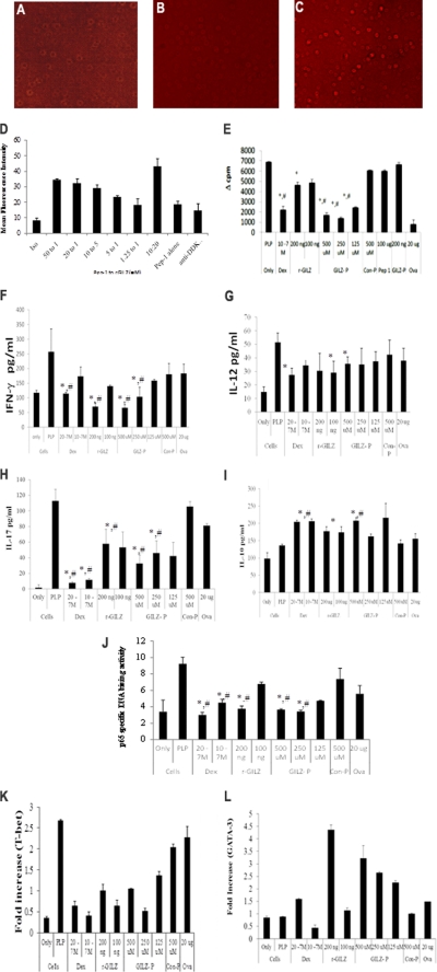
FIGURE 3.
A–D, intracellular delivery of r-GILZ by Pep-1:Jurkat T cells in serum-free HL-1 medium were overlaid with a pre-formed complex of r-GILZ (DDK tag) and Pep-1 at varying concentrations and incubated in a humidified chamber for 1 h at 37 °C. Intracellular delivery of r-GILZ was detected using phosphatidylethanolamine-labeled anti-DDK mAb in permeabilized cells and measured by FACS Calibur. Representative images of cells incubated with (A) r-GILZ alone, (C) 20:1 ratio of Pep-1 to r-GILZ, and (B) pep-1 alone are shown. The delivery efficiency was assessed by mean fluorescence intensity (D). Data are average of two different experiments ± S.E. E–L, treatment with GILZ-peptide suppress T cell responses. CD4+ splenocytes (5 × 10⁵ cells/well) isolated from antigen-primed SJL mice were co-cultured with irradiated syngenic splenocytes as APC and restimulated with PLP139–151 (40 μg/ml) for a total of 72 h (including an 18-h pulse with [3H]thymidine) in the presence of dexamethasone (20−7/10−7 m), r-GILZ (100 and 200 ng), GILZ-P (125–500 μm), and control peptide (C-P)(500 μm) as shown. E, data represent mean Δcpm (cpm of the antigen-stimulated cells − cpm of cells only) ± S.D. from three different experiments. F–I, supernatants collected at 48 h were assessed for IFN-γ (F), IL-12 (G), IL-17 (H), and IL-10 (I) by ELISA. Data are presented as mean ± S.D. J, in separate experiments, draining LNC cultured similarly were harvested at the end of 24 h. Five micrograms of nuclear extracts were tested for binding of the activated p65 NF-κB subunit to an NF-κB consensus sequence using the TransAM NF-κB ELISA kit. The p65 DNA binding activity was calculated as the ratio of absorbance from PLP139–151-stimulated cells to that of unstimulated cells. Values are the average ± S.D. performed 3 times in duplicate. K–L, real time PCR for T-bet (K) and GATA-3 (L) was performed using 50 ng of cDNA isolated from CD4+ LNC of R-EAE mice re-stimulated in vitro with PLP139–151 under the indicated conditions. Treatment with dexamethasone, r-GILZ, and GILZ-peptide down-regulated T-bet (K) and up-regulated GATA-3 (L) expression in activated CD4+ T cells. * and #, p < 0.05 as compared with the cells treated with PLP139–151 alone or cells stimulated in the presence of control-P, respectively.

