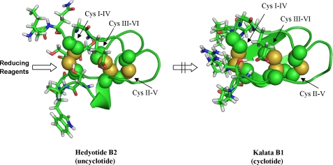
FIGURE 8.
Computer models of hedyotide B2 and kalata B1 (Protein Data Bank code 1NB1). Disulfide bonds (indicated by the arrow) are shown as spheres, and the surrounding amino acids are in stick configuration. For hedyotide B2, its open-ended structure increases the accessibility of Cys I–IV to reducing reagents, whereas for kalata B1 this bond is steric hindered by the cyclic backbone.

