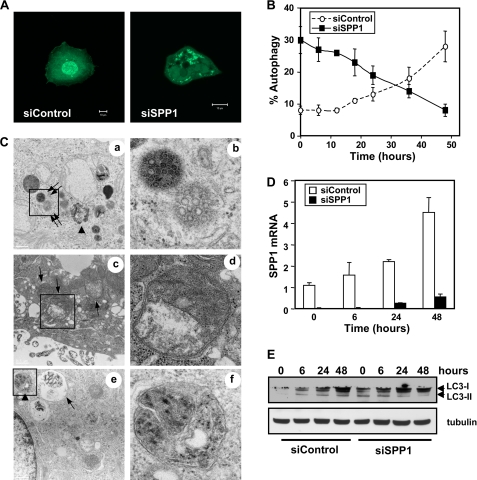
FIGURE 1.
Effect of doxorubicin on autophagy induced by SPP1 depletion. A and B, MCF7 cells were transfected with control scrambled siRNA (siControl) or siRNA targeted to SPP1 (siSPP1) and GFP-LC3 as described under “Experimental Procedures.” Cells were then treated with doxorubicin (1 μg/ml) for 6 h (A) or the indicated times (B and D), and localization of LC3 was examined by confocal microscopy. A, representative images are shown. Scale bars, 10 μm. B, autophagy was quantified. Data are expressed as the percentage of cells showing GFP-LC3 fluorescence in puncta (autophagosome formation) and are means ± S.D. of three independent experiments. At least 100 GFP-LC3-transfected cells were quantified. C–E, MCF7 cells transfected with siControl or siSPP1 were treated with doxorubicin for 6 h (C) or the indicated times (D and E). C, cells were examined by transmission electron microscopy. Typical autophagosomes (arrows), autolysosomes (arrowheads), and multivesicular bodies (double arrows) are boxed and shown at higher magnification. Scale bar, 2 μm (panels a, c, and e) and 0.5 μm (panels b, d, and f). D, SPP1 mRNA levels were determined by quantitative real-time PCR and normalized to glyceraldehyde-3-phosphate dehydrogenase (GAPDH) mRNA. E, cell lysates were analyzed by Western blotting using an anti-LC3 antibody. Blots were reprobed with tubulin to ensure equal loading and transfer.

