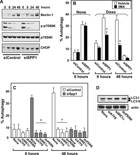
FIGURE 2.
Doxorubicin and ER stress-mediated autophagy. A, MCF7 cells transfected with siControl or siSPP1 were treated with doxorubicin (1 μg/ml) for the indicated times. Equal amounts of lysates were resolved by SDS-PAGE and immunoblotted with the indicated antibodies. Blots were reprobed for total p70S6K to ensure equal loading and transfer. p-p70S6K, phospho-p70S6K. B, MCF7 cells were transfected with siControl or siSPP1 and GFP-LC3. Cells were treated without (None) or with doxorubicin (Doxo) for 6 or 24 h in the absence or presence of 10 mm 3MA, and autophagy was quantified by confocal microscopy. Data are means ± S.D. of three independent experiments. *, p < 0.01 as compared with cells not treated with 3MA. C, MCF7 cells were co-transfected with siControl, siSPP1, siIRE1α, siATF6, or dnPERK and GFP-LC3, as indicated. After treatment with doxorubicin for 6 or 48 h, autophagy was quantified by confocal microscopy. Values are the means ± S.D. of three independent experiments. *, p < 0.01 as compared with None. D, MCF7 cells were co-transfected with siSPP1 and siIRE1α, siATF6, or dnPERK, as indicated, and treated with doxorubicin for 6 h. Cell lysates were analyzed by Western blotting using an anti-LC3 antibody. Blots were reprobed with actin to ensure equal loading and transfer.

