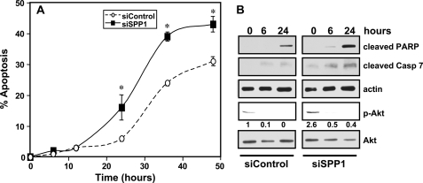
FIGURE 3.
SPP1 depletion sensitizes cells to doxorubicin-induced apoptosis. A and B, MCF7 cells transfected with siControl or siSPP1 were treated with doxorubicin (1 μg/ml) for the indicated times. A, nuclei were stained with Hoechst 33342, and apoptosis was determined by scoring the percentage of cells displaying fragmented, condensed nuclei. At least three fields were analyzed, scoring a minimum of 300 cells. Data are means ± S.D. of three independent experiments. *, p < 0.01 as compared with siControl. B, equal amounts of lysates from duplicate cultures were resolved by SDS-PAGE and immunoblotted with antibodies that recognize cleaved PARP, cleaved caspase 7 (Casp 7), and phospho-Akt (p-Akt). Numbers indicate -fold changes determined by densitometry. Blots were reprobed with anti-actin or anti-Akt to ensure equal loading and transfer.

