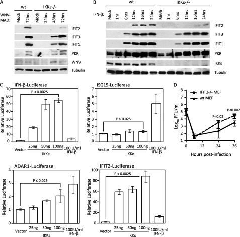
FIGURE 1.
IKKϵ and IFIT2 impose restriction on WNV infection. WT or IKKϵ−/− MEFs were mock-infected or infected with WNV-MAD at an m.o.i. of 1 (A) and mock-stimulated or stimulated with 100 IU/ml IFN-β (B). Protein lysate was collected at the indicated times and immunoblotted using IFIT2, IFIT3, IFIT1, PKR, and IKKϵ antibodies. Tubulin was used as loading control. C, HEK293 cells were cotransfected with pCMV-Renilla and either pIFN-β-Luciferase (top left panel), pISG15-Luciferase (top right panel), pADAR1-Luciferase (bottom left panel), or pIFIT2-Luciferase (bottom right panel). 16 h later, cells were either mock-stimulated (vector cotransfection); stimulated by transfection with 25 ng, 50 ng, or 100 ng of an IKKϵ expression plasmid; or treated with 100 IU/ml IFN-β. Cells were harvested 48 h post-transfection, and luciferase expression was measured and normalized to Renilla. The relative luciferase value was calculated as fold induction over induction of the vector that was set to 1. Statistical analysis was performed with Student's t test. D, WT or IFIT2−/− MEFs were infected with WNV-MAD at an m.o.i. of 5. At the indicated time points post-infection, culture supernatants were collected, and virus titers were determined by plaque assay on BHK cells. C and D, data are mean ± S.D. of three independent experiments performed in triplicate. p values were calculated using Student's t test to determine statistical significance.

