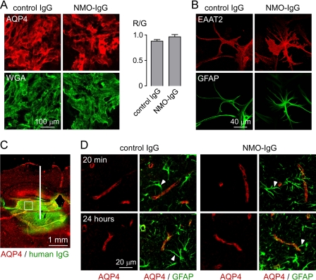
FIGURE 7.
Perivascular astrocyte membrane localization of AQP4 in vivo following intracerebral injection of purified IgG from NMO sera. A, left, mouse astrocytes stained for surface AQP4 (red) and plasma membrane (WGA, green) after a 24-h incubation with purified IgG from NMO or control sera (done as in Fig. 2). Right, quantification of surface AQP4 (mean ± S.E. (error bars), n = 10). B, EAAT2 (red) and GFAP (green) staining of astrocytes after 24-h incubation with control or NMO-IgG. C, intracerebral injection. Brain section stained for human IgG (green, showing area of diffusion) and AQP4 (red). White line, needle tract; white square, area where AQP4 localization was assessed in D. D, confocal micrographs of AQP4 (red) and GFAP (green) at 20 min and 24 h after injection of control or NMO-IgG. Arrowheads indicate GFAP-stained astrocyte foot processes. Results are representative of micrographs from 2–4 mice per condition.

