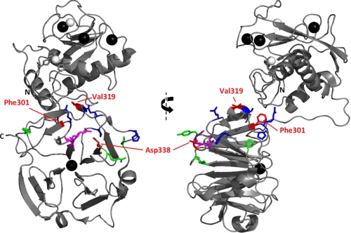
FIGURE 5.
Residues of HPX-1 implicated in collagen binding by mutagenesis and assay. Orthogonal views of the crystal structure of MMP-1* are shown as a ribbon diagram with the left-hand side in the same orientation as that in Fig. 3A. Mutated HPX-1 residues are shown as sticks and colored according to the collagen binding activity of the mutant, i.e. Phe301, Val319, and Asp338 in red (Kd ≥ 24 μm); Tyr309, Arg337, and Pro361 in green (16 μm < Kd < 24 μm); and Arg300, Ser318, Pro325, Gln354, and His358 in blue (Kd ≤ 16 μm). Residues Ile290 and Arg291, previously identified as an exosite (29), are shown as magenta sticks.

