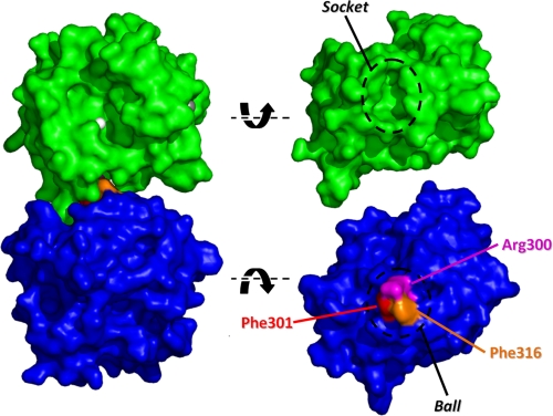
FIGURE 7.
The ball and socket joint in the CAT-HPX interface of MMP-1*. A space-filling model of the crystal structure of MMP-1* is shown looking into the active site cleft with the Zn2+ ion colored white. Dislocation of the ball and socket joint and rotation of the CAT (green) and HPX (blue) domains reveals the interacting surfaces. The three residues comprising the ball are highlighted.

